by Andrew I Spielman
How to cite this page: Spielman, AI. History of Restorative and Esthetic Dentistry. In. Illustrated Encyclopedia of the History of Dentistry. 2023. https://historyofdentistryandmedicine.com/
History of restorative materials, agents, and tools
The first evidence of tooth restoration is from 11,000 BCE, Upper Paleolithic. In dental specimens found in Tuscany, Italy, the exposed pulp chamber of two anterior teeth has been enlarged and restored using bitumen, vegetable fibers, and probably the hair. The most likely tools used to enlarge the canals were flint stones (1). More recent evidence of restorative intervention using beeswax comes from4500 BCE. The Lonche jaw, found in Slovenia, showed a lower left canine had traces of restoration (2, Bernardini).
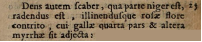
There is a gap in our knowledge regarding restorative dentistry in ancient Egypt or Greece. Celsus, the first-century Roman physician, however, recommends scraping the black portion (parte niger) from the cavity and filling the tooth with a mixture of rose leaves (rosae flore), nutgalls (cui gallae) and myrrh (myrrhae). (See image from his 1687 edition of De Medicina on the left (3)

The subsequent significant development in the history of tooth restorations occurred in 1460 when Giovanni d’Arcoli (1412-1484), a Professor at the University of Bologna, first used gold foil to fill teeth (4). Giovanni da Vigo (1460-1520) of Rapallo, personal physician to Pope Julius II in 1514, goes one step further, cleaning carious teeth (corrosio) before filling them with gold foil, using drills, files, and scrapers (trapano, lima, scalpo) (5, 6). (See image from the text by d’Arcoli, on the right).
One had to understand the normal appearance, structure, and function to restore teeth. The first dedicated study on dental problems (Artzneybuch) was published in 1530 in German and had seen 11 editions (7). It was part of a medicinal book to treat “all kinds of ailments….from head to toe” and directed to itinerant healers and tooth drawers. The first scientific work on teeth was that of the Spanish, Francesco Martinez’s text in 1557 entitled Coloquio Breve, and six years later of the Italian Bartolomeo Eustachio 1563 publication, Libellus de dentibus (8). Soon other works dedicated to teeth appeared in France, Martin Bernardin, 1679, and in England, Charles Allen, 1687. These books set the stage for more detailed studies on diseases of the teeth and their restoration.
Although known from the 7th century, amalgam appeared as a restorative material in 1528 when Johannes Stockerius (1453-1513), a German physician, recommended it. A significant advancement in techniques, instruments, and approaches to dentistry comes from Ambroise Paré (1510? -1590), one of the most outstanding surgeons of the Renaissance. A practicing barber-surgeon, he designed new surgical instruments, extraction tools, obturators, gentle wound healing, and arterial ligation techniques and described restorative approaches in his book entitled Oeuvres (Works) in1575. Paré still believed in the tooth worm and suggested applying something caustic, hot, dissolved in vinegar or theriac (9). In that same spirit of using a caustic agent, Johann Heurnius (1543-1601), a Dutch professor of medicine at the University of Leiden, suggests using sulphuric oil (oleum sulphuris) to destroy the dental pulp and to reduce the roughness and sharp edges on teeth (10). An alternative to gold restorations comes from Jacques Guillemeau (1550–1613), a French surgeon from Orleans who, in 1612, used a mineral paste made of mastic and coral powder (11).
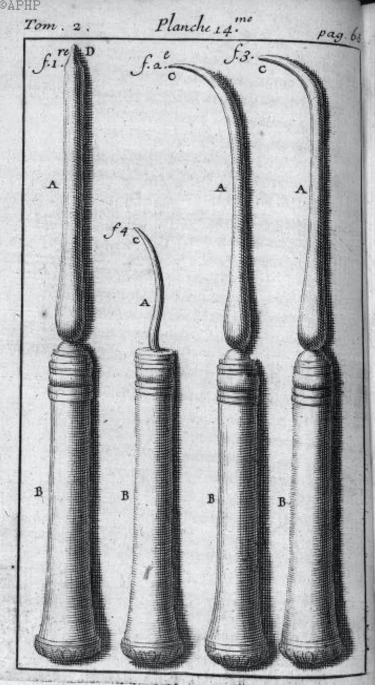
Compared to gold or mineral paste, a cheaper and more reliable alternative was lead or tin. Pierre Fauchard (1678-1761), the “Father of dentistry,” in his 1728 work Le Chirurgien Dentist displayed materials and tools that he employed in restorative dentistry (see Figure on the left, Fauchard, Planche 14, p.63, Vol 2, 1746). Fauchard used files, an Archimedes-type drill, and sharp instruments to enlarge a cavity and remove decay. He cauterized the cavity with a hot tool and filled it with lead or tin. The French word for lead and a dental filling is the same, plombe.
In 1796, Frederich Hirsch (1750-1827), a German court dentist from Göttingen, published the first cement-like restorative material made of quicklime (calcium oxide) (12). It was not a particularly successful material. Forty-some years later, in 1840, Wolfsohn of Berlin developed a quick-setting restorative material made of a mixture of Sandarac, a resin from a small cypress-like tree, chalk, and mastic mixed with ether (13). However, the restoration required frequent replacements because of its foul odor.
During the 19th century, when more frequent dental appointments required a temporary restoration, there was the need for a material that would not harden too quickly or could be removed at the next appointment. Gutta-percha which debuted in the middle of the 19th century, appeared to fit the purpose. Its name, from two Malay words: Getah (gum) and Pertja (name of the gum tree), was derived from trees primarily from Malaysia and Indonesia. Isolated in 1842, gutta-percha was first used for surgical splints. In 1847, Edwin Truman used it as a temporary restorative material. It also found application as a denture base material for a short while. However, Jonathan Taft of Michigan suggested its longer-lasting use in root canal therapy in 1859 (14).
As demand increased for better restorative materials, in 1840, Charles Sylvester and A. Rostaing, a father-son team from Dresden, Germany, developed a new cement containing zinc oxide and phosphoric acid. The secret formula of the Cäment was never revealed (15). A much more advanced cement also came from Germany, where in 1892, Otto Hoffmann developed a high-quality zinc phosphate cement. It had a monopoly on the market until World War I. Parallel with the development of dental cement, there was a need for an esthetic restorative material for anterior teeth. In 1878 the “translucent cement” of Thomas Fletcher in London was patented (16). A more successful and esthetically pleasing cement was formulated by Paul Steenbock, a Berlin chemist, in 1903. It contained zinc oxysulfate cement, aluminum beryllium, calcium oxide, and silicic acid.
Because acidic components of several restorative materials caused pulp damage chemically or by transmitted heat, cavity liners were developed. A layer of asbestos under the restorative material was included in 1846 by James Robinson of London (17), and in 1930, Hermann BW proposed calcium hydroxide to protect the dental pulp. Since then, zinc oxide eugenol, glass ionomers, and resin-based liners have been employed. (18)
Eugenol and isoeugenol are extracted from cloves and nutmeg, respectively. Both have been used to reduce dental pain and pulp inflammation. Zinc oxide eugenol was used as part of the temporary restoration for the better part of the 20th century. Nutmeg, which contains clove oil, among many herbal medicines in the 17th century, was considered powerful enough to ward off the plague. It was highly prized as a spice and source of herbal remedies. The Dutch, who dominated the spice trade in the 17th century, based on the Treaty of Breda in 1667, traded the island of Manhattan to the British in return for the spice island of Run, rich in nutmeg. Nutmeg is the reason Manhattan speaks English today and not Dutch.
Composite bonding agent was introduced to dentistry in 1955. It started with the Swiss chemist Hagger at the DeTrey/Amalgamated Dental Company with a dentin bonding agent, but Michael Buonocore (19) that 1955 established the field of acid etch technique for bonding to both dentin and enamel. Since its introduction, several generations of bonding agents have emerged, from no-etch to total-etch to self-etch, with constantly improving qualities. Today’s composite agents are dentin-adhesive with either an etch and rinse or a self-etch system (20). They are aesthetically pleasing, enduring, and placed on the anterior and posterior teeth equally successfully.
Amalgam has been the focus of dentistry for at least 200 years, and it deserves a separate section. The first mention of amalgam is in 659 CE as silver dough in Su Kung’s Materia Medica, a compendium for healing in ancient China.
It resurfaced in several Chinese publications over the following centuries, including The Essentials of Materia Medica, in 1505. Its composition is also displayed: 100 parts mercury, 45 parts silver, and 900 parts tin (21,22).
Amalgam used specifically for dental filling appears in a prescription of Praxis, a 1528 work of Johannes Stockerius (1453-1513), a German physician (23). In it Stockerius states: Fac canalem de auro fino, et per canalem cauterizetur dens cum alio aureo usque ad mortificationem radicis dentis, postea imple foramen cum Amalgama, facta ex Vitriolo et Mercurio ita ut sequitur. Rx. vitrioli, et dissolve cum aceto forti in patella, adde Mercuri quantum uis. Deinde decoquatur, et convertetur Mercurius in Amalgama, et illud Amalgama pone in foramen dentis, et durescit sicut lapis, et valet in omni foramine. Translation from Hoffmann-Axthelm: Make a tube from refined gold, and the tooth should be cauterized through this with another gold (instrument) until the root is deadened; after that, fill the hole with an amalgam compounded from vitriol and mercury as follows: dissolve vitriol with a strong acid in a bowl, and add sufficient amount of mercury to it. Then it should be boiled, and the mercury will transform into an amalgam; put this amalgam into the hole of the tooth, and it will become as hard as a stone and remain firmly in any hole.
The first amalgam restoration, in situ, was discovered in a grave dated to 1601 in Crailsheim, north of Munich. The remains of Princess Anna Ursula of Brunswick and Luneburg, an ancestor of the House of Hannover and England, showed a molar restoration with gold foil and amalgam. (24).
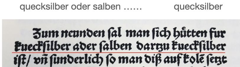
The dangers of mercury (argentum vivum) used to treat syphilis have been well-known since the early 16th century. Zene Artzney, the first dental book published in 1530, cautions about “quicksilver” or mercury. It states, “Beware of quicksilver or salves in which there is quicksilver” (6). (See image on the left). In 1606, Peter van Foreest warned about the effect of mercury on teeth. He states: Nam argentum vivum non minus quam cætera dentibus infestum est: In fact, mercury is dangerous to the rest of the teeth. (25)
Sir Isaac Newton (1642-1727), among his many accomplishments, 1686 developed a low-melting fusible metal alloy containing eight parts bismuth: five parts lead: and three parts tin. That alloy served as the basis of dental amalgam 132 years later. First, in 1775 Jean d’Arcet, a chemist and director of the Sèvres Porcelain manufacture, proposed a mixture of bismuth: tin: lead in a 5:3:2 ratio for porcelain manufacturing using Newton’s alloy. Subsequently, in 1818 Louis Nicolas Reignart (1780-1847) suggested adding a mixture of mercury at a ratio of 1:10 to create dental amalgam. Upon heating, it became malleable and placed in small increments into the dental cavity, and one could shape it into the desired form. (26). Further modification to the composition by adding cadmium, copper, pulverized silver, and coin shavings further altered the handling property of amalgam. During the next decade (1826-1835), August Onesime Taveau tinkers with its composition using pulverized silver and mercury, creating a silver paste. In 1848 Thomas Evans of Philadelphia, working in Paris, used a modified ratio of cadmium: tin: silver 1:3:1 ratio to improve it. Finally, in 1855 Elisha Townsend of Philadelphia suggested a 4:5 silver: tin ratio for amalgam, a composition that has endured for decades.
There was a unique period in American dentistry related to amalgam called The Amalgam War. It had to do with Edward and Moses Crawcours, uncle and nephew team of London, making a trip to the East coast of America in 1833 using silver coin shavings instead of pulverized silver to create their “secret” mix of Royal Mineral Succedaneum. Amalgam was received with reservation by some practitioners primarily because the Crawcours, two untrained dentists, became financially successful and siphoned away many patients. Opposition among local dentists grew, leading to a campaign to ban amalgam and send the Crawcours back to Europe. The ban on using amalgam was lifted around 1850.
Cavity preparation was a particular point in the history of restorative dentistry, particularly for amalgam restorations. The most crucial individual that stands out in its development is Greene Vardiman Black, a dentist, and educator at the end of the 19th and beginning of the 20th century. His 1908 book Operative Dentistry and the principle of extension for prevention incavity preparation dominated most of the 20th century (27).
Inlays have a relatively short history. It appeared in 1834 in Joseph Calmann Linderer’s work Lehre von den gesammten Zahnoperation (Instructions for all dental operations) (28). Linderer termed a “veneer filling, ” a walrus bone round peg forced into a cavity using a small hammer and retained with fish-glue cement. The first porcelain inlay was the work of Edward Maynard of Washington in 1857 (29). He cut shapes from porcelain teeth that fit a precisely prepared cavity and secured it in place with gold foil. Subsequently, the inlay was secured with better-adhering gutta-percha or cement. The gold inlay became a method of choice only after the precise casting of gold using the centrifugal casting machine, invented by William H. Taggart, was achieved in 1907 (30). The wax model created in situ or based on an impression was embedded and cast in gold. The technique, literally and figuratively, was the “gold” standard of inlay preparation until less technique-sensitive and cheaper methods took over later in the 20th century.
- 1. Oxilia (2017)
- 2. Bernardini
- 3. Celsus p. 444
- 4. D’Arcoli p.140
- 5. da Vigo p.533
- 6. Zene Artzney
- 7. Artzneybuch
- 8. Eustachio
- 9. Paré p.612
- 10. Heurni p.61
- 11. Guillemeau (a). p.502-3
- 12. Hirsch p.131-2
- 13. Nessel p.237
- 14 Taft (a)
- 15. Hoffmann-Axthelm (a). p. 294
- 16. Fletcher 1878
- 17. Robinson
- 18. Hermann
- 19. Buonocore
- 20. Sofan
- 21. Chu p.553
- 22. Hoffmann-Axthelm (b). p.43
- 23. ibid p.156
- 24. Czarnetzki
- 25. Foreest p.214-217
- 26. Reignart
- 27. Black p.105-210
- 28. Linderer
- 29. Wheeler p.547
- 30. Taggart (a) (b)
Mechanical removal of decayed tooth structure was an essential component of restorative dentistry and defined the profession for the better part of its existence. Only in the last two decades has minimally invasive dentistry, and for the past 75 years, preventive, non-invasive dentistry has become a new direction. But restorative dentistry was associated with drilling for the better part of its modern existence.
The first evidence of mechanical removal of decayed tooth structure comes from 7000 BCE. During the excavation of ancient graves in Pakistan in 1995, several teeth with traces of drilling, probably with a stone flint, came to light. (1) Trephines, sometimes using a hot iron to open up the skull or drain purulent teeth, were used in Ancient Egypt. Evidence of new bone deposits around such charred openings indicates that the subjects survived the ordeal. The primitive drills were turned by holding the shaft between two hands and moving the palms back and forth. Alternatively, a string or bow drill was used. The Greeks and Romans were familiar with the string and bow drill and opened teeth using a trephine (2).
Giovanni d’Arcoli, the first to restore teeth with gold foil, most likely used such a drill. Giovanni da Vigo (1460-1520) of Rapallo, personal physician to Pope Julius II, 1514 goes one step further, cleaning carious teeth (corrosio) before filling them with gold foil, using a string drill, files, and scrapers (trapano, lima, scalpo) (3,4). A section of the first dental book meant for itinerant tooth drawers, Zene Artzney, describes using the drill and scrapers in 1530. One of the first illustrations of the string drill in medieval texts appears in Andrea dela Croce’s (1500-1575) illustrated Chirurgiæ, published in 1573 (5).

Drille de bijoutier (jeweler’s drill) as seen in Andrea dela Croce, 1573, p.41 (https://commons.wikimedia.org/wikimedia).
Roughly a century later, Cornelius Solingen (1641-1687) used a hand drill to smooth the rough edges of the carious lesions (6). An illustration and more detailed description of the bow drill appears in Pierre Fauchard’s 1746 second edition. However, the drill is almost exclusively used to drill holes into the denture base made of bone (7). Fauchard’s drill was too bulky to be used inside the mouth. In addition, Fauchard used files, rasps, and hand scrapers to remove the carious lesion from the tooth, but not drills.
The french author, Joyurdain, Anselme-Louis-Bernard-Brechillet (1734-1816), Nouveaux élémens d’odontalgie, Paris, 1756 (8, p.215) describes what is by all account the first drill (porte ecarissoir).

A significant advance in the history of cavity preparation came from John Greenwood (1760-1810), one of George Washington’s favorite dentists. In 1790, Greenwood modified his mother-in-law’s foot-treadle spinning wheel and created the first foot-operated dental drill.
Dental drill belonging to John Greenwood ( image on the left). Currently at the New York Academy of Medicine, where the drill has been located since 1934. Image reproduced with permission.
In 1803, a small handheld drill is devised by the Berlin dentist Heinrich Lautenschläger (9) (image to the left). It is a hand-cranked drill small enough to allow its use inside the oral cavity. The drill was not used to remove the carious lesion from the crown, only to enlarge the root canal to insert a pin to anchor the crown. Such intervention preceded formal root canal treatment to be introduced only in the middle of the 19th century (see History of Endodontics).
The importance of future dental drills was not recognized in 1829 when James Nasmyth, a Scottish engineer, invented the flexible shaft and coiled spring to rotate a drill (“suitably close-coiled spiral steel wire conveyed rotary motion”). That invention found a ready application in dental drill designs thirty years later. (10, 11).
One of the more inventive practitioners of the early 19th century was Charles F. Maury, a Parisian dentist that introduced a hand drill operated by a plunger (porte- forêt) activating a small bur via a cord. It could reach any tooth like a contra-angle drill (14).

A decade later, in 1838, John Lewis of Burlington, Vermont, patented a dental drill that he called a universal bevel gear. (15). Several additional drills were introduced in the 19th century by the French Pierre Joachim LeFoulon (1841) and the British George Fellows Harrington (1864), a spring-operated drill that required wounding up, see image) or the American Josiah Foster Flagg (1846), John A. Chevallier (1850), and Charles Merry (1858). These drills required one or two hands, limiting the dentist’s holding a mirror or retracting the cheek.

Image to the left – Harrington clockwork drill (1864) (Harrington’s Clockwork Dental Drill, the “Erado”, n.d. Artifacts. Harrington’s Clockwork Dental Drill, the “Erado”, [Co102130]. (Image on the left). https://jstor.org/stable/community.26293170).

The breakthrough came in 1871 with the invention of the patented foot-pedal-operated dental drill. James Beall Morrison (1829-1917) was an Ohio dentist whose dental engine was based on Greenwood’s 1790 foot-treadle spinning wheel. It had a 2000 rpm speed and gave the dentist two hands to operate (16). In 1883, a foot pedal-operated dental drill suspended from the ceiling made drilling more convenient (see image on the left, from Taft, 1883, 3rd edition).
Building on Edison’s new invention of electricity, starting in 1872, George F. Green, a mechanic at the S.S. White Co in Philadelphia, demonstrated an electric (battery-operated) dental drill. Further improvements by Erwin Moritz Reiniger (1854-1909), a mechanical engineer at the University of Erlangen in 1887 (17), have made the drill usable.

The drill got a new configuration with the foot pedal activating an electric engine instead of a pumping motion. One could hang the electric motor from the wall and connect the engine to the drill via a belt or cord. Oscar H Pieper patented such a wall-mounted electric dental drill in 1893 (18, image to the right). It had speeds from 250 to 6000 rpm.

In 1909, a hand-held dental drill with a small battery-operated electric engine attached to it was filed by Howard A Whiteside of New York and patented in 1916. (19). Whiteside had several patents to his name over the next two decades, most of which improved his dental engines.
Still, dentists’ broad acceptance of the expensive electric drill occurred only at the beginning of the 20c., when the electric drill was incorporated into the adjustable dental unit and chair. By 1917, the Ritter Company of Rochester, NY, combined the various components, drill, electric motor, tray, water spray, cuspidor, etc., into a functional dental unit (see the section on the history of the dental chair).
At the end of the 19th century, mechanical handpieces, straight and contra-angle, were introduced, and steel drills were replaced with diamonds. By 1936 the speed of the cord-driven dental drill was improved from 6,000 to 24,000 rpm. Nevertheless, considerable vibration was still transmitted into the tooth of the sitting patient.
The next significant advance in the evolution of the dental drill occurred when the high-speed turbine, water- and later, air-driven, were introduced in 1957. The development of the turbine had input from several inventors: Ivar Norlen of Sweden in 1948, the air turbine developed by John Victor Borden originally in 1946, and commercially as the “Airotor” in 1957. Because of the heat generated at such speed and the danger of damaging the viable tooth, a continuous water cooling system was attached. It could have 150-300,000 rpm speeds and required a constant water-cooling system. Today high-speed dental units can run up to 800.000 rpm and are the main tool to remove enamel structure. However, as less-invasive or minimally invasive procedures are being introduced into dentistry, not to mention that regeneration of tooth structures is emerging, the role of the drill will become part of history.
History of the dental bur used in drills

The rotating sharp tools to create a hole in skull bone go back to the Late Neolithic Ages, 7000 B.C.E., and were widely used in ancient civilizations (20). Burs were perfected for bone or metal cutting throughout the centuries. During the 15 and 16th centuries, skull trepanation became a standard practice to treat mental ailments or “remove the Stone of Madness” (Painting by Hieronymus Bosch, 1501-1505, Museo National El Prado, Madrid (Image to the right).

In his 1570 Works (Oeuvres), Ambroise Pare displays several hand drills and burs for the procedure (see image to the left). These burs were hand-made using cast iron. Once mass production of steel was improved by the Bessemer Process in 1855, steel burs became common and cheaper to produce.
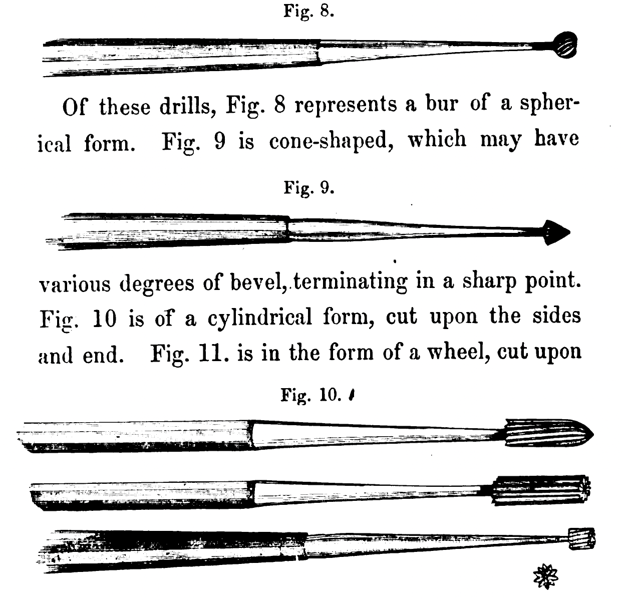
In an 1859 dental school textbook, Jonathan Taft dedicates considerable time to the description of hand-held dental burs (21) (see image on the right). When Taft’s 3d edition was published in 1883, burs were attached to foot-pedal-operated belt-driven mechanical devices. (22). These burs were machine-made to ensure consistency and efficiency. As steel-making was improved via the Siemens-Martin process over the next century and carbide of tungsten, titanium, and tantalum were forged into steel, such burs could be used in high-speed dental cutting instruments, the state-of-the-art after its introduction in 1957.
History of restorative instruments

In the annals of dentistry, restoring teeth was a relatively late addition. Solutions to address dental pain appeared in ancient Roman works. The first systematic approach to using instruments dedicated to tooth restoration occurs in the 8-9th century Arab texts. The first instruments were sharp cauterization tools (23, 24) used to scrape the bottom of a large cavity or cauterize a painful tooth, killing the nerve. Arab physicians even developed a funnel to protect burning the soft tissue while cauterizing the tooth. (25)
These early instruments were primarily for removal or stopping caries progress, but not necessarily filling the cavity. Only during the 16th century did we see tools to plug a tooth. The primary texts of the 16th century were from barber-surgeons . Still, most of the instruments displayed in the work of Ambroise Paré, or Scultetus, were forceps, pelicans, and elevators, surgical in nature.
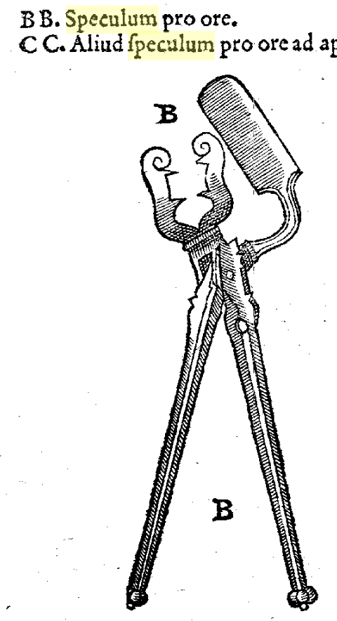
Guy de Chauliac, a French surgeon, was the first to describe a retractor combined with a dental mirror (speculum pro ore). Its illustration first appears only in the 1585 edition (26, figure on the right). The mirror was a flat, highly polished metal surface with a retractor. Flat shiny bras mirror was well known to ancient Egyptians, Greeks, and Romans. During the Renaissance, glass mirrors coated with tin-mercury amalgam were manufactured in Venice. The modern mirror with silver-impregnated glass was invented in 1835 by Justus von Liebig.
Although a brief mention of using a dental mirror appears in 1811 (12), the first full description of the modern dental mirror comes from James Snell (1831) (13), a London Dentist. He describes but does not illustrate small concave intraoral glass and metal mirrors custom-made for him to visualize cavities and calculus on the invisible side of the teeth.

Building on that invention, the ever resourceful C.F.J. Maury, the dentist of the l’Ecole Royale Polytechnique, and inventor of the porte-forêt dental drill, also illustrates the modern dental mirror, miroire du dentiste (image on the left) (14). Some studies incorrectly attribute the discovery of the dental mirror to Bartholomew Ruspini. The latter described an extra-oral mirror for patients to see themselves, rather than the one used intraorally by dentists.
Many of the ornate, ivory handle oversized oval intraoral mirrors were replaced by metal, round, flat, or concave mirrors in the middle of the 19th century. Once sterilization of dental instruments was a regular practice starting with the beginning of the 20th century, stainless-steel, corrosion-resistant metal handle intraoral mirrors replaced them.

The dental probe (explorer) appears in the work of the French surgeon, Garengeot. In 1727 in his second edition on dental and surgical instruments: Nouveau traité des instrumens de chirurgie les plus utiles et de plusieurs nouvelles machines propres pour les maladies des os, for the first time we see a dental probe (sonde) depicted (27, image on the right).

The carious substance was removed with instruments borrowed from other professions: a chisel, knife, file, or a sharp scraper. Goldsmiths of the 17th century used small scrapers (28). Adjusting them to fit the opening of a typical tooth was done intuitively, and the process was named interchangeably as scraping, excavating, scooping, or cutting. Scraping the bottom of a carious lesion was recommended as early as 1530 when Zene Artzney, the first book entirely dedicated to dentistry, was published(4). Subsequent books published in the 16th century by Francisco Martinez de Castrillo (29) included sharp instruments and an early form of an excavator (see image on the right) to scrape the cavity. Pare (30) recommends cauterization with a small flat hot instrument, much like the 9th-century Arab physicians recommended, but spent little time reshaping the cavity or restoring it.
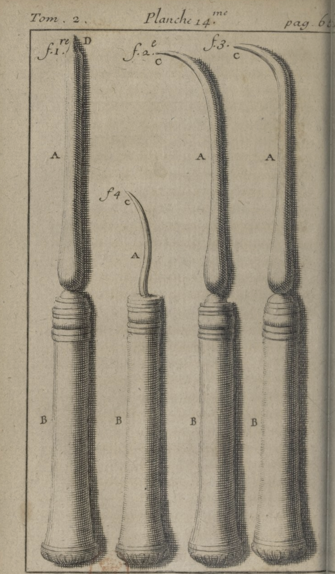
Instruments to remove caries gained importance and variety as dentistry entered a restorative phase at the beginning of the 18th century. Pierre Fauchard depicts four different scrapers/excavators (image on the left) in his 1728 seminal book (31).
In 1841, Pierre Joachim Lefoulon, a French dentist, described excavators (curette) and hand drills for removing caries in the absence of a reliable drill to remove soft dentin. He recommended tin, gold, or platinum sheets packed into the cavity using special pluggers, and when completed, he burnished their edge.
In the 19th and early 20th centuries, several turning points in developing restorative instruments are important. The first was the move from handcrafted, ivory, or metal-made individualized instruments to commercially available mass-marketed stainless steel instruments. This switch occurred toward the middle of the 19th century with the establishment of dental suppliers such as S.S. White in Philadelphia (1844) and Ash and Sons in London (1820). As demand for services increased, so did the available range of hand instruments for excavation, shaping, and restoring cavities. These included an ever-growing array of instruments such as probes, scrapers, enamel cutters, chisels, excavators, stoppers, pluggers, and burnishers. Handsets of over 150 instruments were not uncommon.
The next major shift in instrument design came at the end of the 19th century when the microbial nature of diseases urged the sterilization of dental instruments. This required instruments that could withstand chemical and thermal exposure during sterilization.
Finally, with the appearance of dental schools and dental education in the second half of the 19th century, clinical practice entered a more formal process of developing, categorizing, standardizing, and streamlining the number and types of instruments used for restorative dentistry. This culminated with the publication in 1908 of the two-volume Operative Dentistry text of G.W. Black (32). Black’s methods defined restorative and operative dentistry for a century.
Today’s array of instruments is extremely diverse and highly specialized. However, as dentistry is moving toward less and less invasive interventions and more digitized approaches, many instruments are either discarded or less used. For instance, the cautery is no longer in use; the excavator is less and less used, the mirror is being supplemented by intraoral cameras, and the probe is no longer recommended to detect a carious lesion to avoid more damage.
- Coppa
- Hoffmann-Axthelm (c). p.299.
- da Vigo p.533
- Zene Artzney
- Croce (a) p.41, idem (b)
- Solingen p.119
- 7. Fauchard (b) v. II, p.236.
- Jourdain, 2015
- Lautenschlager p.55
- Wilson p.34
- Nasmyth, p. 442
- Murphy p.75
- Snell p.129
- Maury (a), Planche IV; Maury (b). Planche 23-24
- Lewis
- Morrison
- Hoffmann-Axthelm (d). p.308
- Pieper,
- Whiteside
- Papagrigorakis
- Taft (a) p.100
- Taft (b) p.113
- Albucasis
- Avicenna p.
- Sharaf al-Din, p.29
- Guy de Chauliac p. 656.
- Garengeot, p.42
- Badcock, p.1
- Martinez de Castrillo p.48
- Pare p.395D
- Fauchard (a) v. II p.65
- Black
History of tooth whitening and bleaching agents
Tooth whitening and bleaching agents have different purposes. The former removes extrinsic; the latter neutralizes intrinsic pigmentation. Tooth whitening uses abrasive agents; bleaching involves free radicals. For a good review of the history and current clinical aspects, consult reference (1).
Tooth whitening goes back to Rhazes (854-925/932?), a Persian erudite, philosopher, polymath, and physician of the 9th century. Rhazes’ 23-volume Kitab al-Hawi has a section on tooth preservation which includes a mixture for a brightening dentifrice containing birthwort, ocean crab, mussel ashes, soda borax, salt burnt with honey, juniper incense, pumice, glass, burnt thyme and mugwort (2).
Medieval medical literature often has a section on the cure for blackened teeth (dentes nigros or ad dealbantos dentes). Starting with the mid-16th century, Johann Craton von Kraftheim (1519-1586) recommended Oleum vitrioli (or ethyl ether) for tooth whitening (3). Ether oil was known since 1540 and described by Hyeronimus Cardanus (4). In 1606, Peter van Foreest, a.k.a. the Dutch Hippocrates, recommends a dentifrice for tooth whitening that contains hyssop, oregano, menthae, alum, pulverized dear stag, and table salt, all reduced to ash, mixed with pepper, chrysanthemum, mastic, sweet myrrh, and cinnamon (5). In a 1623 work, the same author lists five more mixtures for tooth whitening containing abrasive agents like pulverized bovine, dear stag and sepia (cuttlefish) bone, sponge stone and pumice, table salt, red or white coral; stabilizing agents like acacia (Arabic) gum, mastic or myrrh and active ingredients to mask halitosis including nutmeg (source of isoeugenol), “aqua menthae” (from dried leaves of menthae), cinnamon, and solvents that include white vine and rose water. (6). These recipes contained many active ingredients in today’s tooth whiteners: multiple abrasive and whitening agents. At the time, although esthetic concerns were not common, having black teeth must have been an issue for some. This became particularly concerning when sugar became available in the 16th century. Many who could afford to buy sugar have experienced an increase in blackened teeth. A good example was Elisabeth I, queen of England, who became so addicted to sugar that contemporary complained about her mood swings, black teeth, small pursed lips to hide it, and her tendency to cover the mirrors in her palace to avoid seeing her unappealing self-image.
In 1619, Rodericus a Fonseca, a Portuguese physician, suggested white bread, red corral, and powdered alabaster mixed with pulverized rosemary leaves (Folia rosmarini) used as a dentifrice to whiten teeth (6). Many of the 16-17th century dentifrice recipes were compiled by Thomas Burnet in a 1697 work entitled Thesaurus medicinæ practicæ (6).
By the beginning of the 19th century, awareness of one’s appearance and oral hygiene routines became more common. A period text recommending at-home prepared tooth whitener included lemon juice mixed with burnt alum and salt (7).
The first textbook on oral hygiene was written by Levy Spear Parmly (1818), the Father of Oral Hygiene. The book entitled A Practical Guide to the Management of the Teeth provided a detailed description of the tools for oral hygiene. Oral cleaning became an integral part of what dental offices offered. As a result, demand from patients for stain removal increased. Today’s tooth whitening agents are abrasive but less damaging than those recommended in the 16 through 19th centuries. New tooth whiteners include silicon dioxide, pumice of various coarseness, calcium carbonate, and polishing agents like tin oxide, corundum, or diamond particles containing paste.
Chlorinated lime (calcium hypochlorite) combined with sodium chloride as a bleaching agent was first reported in 1848 at the American Society of Dental Surgeons meeting (8). The topic of tooth discoloration frequently appeared in period publications. Another strong oxidizing agent, sodium hypochlorite (Labarraque Solution), was reported in 1861 (9). Antoine Germain Labarraque developed a strong antiseptic solution of sodium and calcium hypochlorite in 1825, widely used in disinfecting dead animals and subsequently used by Semmelweis in 1846. In 1865 James Truman (10,11) sealed into tooth cotton with a mixture of strong acid and chloride lime and let patients walk home and return for the agent to be changed several times weekly, thus initiating the technique later dubbed “the walking bleach”. Concentrated oxalic acid, placed in the pulp chamber for 3-6 minutes, appeared to bleach teeth significantly (12).
Hydrogen peroxide was first suggested by Harlan in 1884. (13). Several minutes of exposure to concentrated H2O2 was reported as an effective bleaching method. Harlan’s method seems to have been upheld for the past century. In 1961 sodium perborate and water were placed into the pulp chamber of discolored non-vital teeth. Subsequently, 25% (Pyrozone) and 30% (Superoxone) hydrogen peroxide in ether replaced water in the solution, achieving even better results (14). Carbamide, another strong bleaching agent, was introduced in the 1960s due to a chance observation of tooth whitening following 10% carbamide peroxide treatment for post-orthodontic gingivitis. That observation led to the rise of vital-tooth bleaching techniques using trays and the night-guard vital bleaching technique (15). Home-bleaching products using 10% carbamide-peroxide became available in 1989.
When tooth whitening or bleaching failed to improve esthetics, porcelain veneers were applied. Introduced by Charles Pincus in California in the 1930s (16), he placed thin porcelain veneers on front teeth to improve the appearance of Hollywood stars. The method, though did not take off because the adhesion of the veneers was poor. (17). Only when better etching and bonding between the silane (SiO2) groups of the porcelain veneer and the bisphenol glycidyl methacrylate (bis-GMA), appear in the late 1970s did porcelain laminates became a more reliable option for esthetic treatment (18).
- 1. Kwon
- 2. Hoffmann-Axthelm (e). p.92
- 3. Burnet p.273
- 4. Gessner p.425, 428
- 5. Foreest p.214-217
- 6. Burnet p.273
- 7. Woodforde (d). p. 43
- 8. Dwinelle p.57-61
- 9. Woodnut p.662
- 10. Truman (a) p.70
- 11. Truman (b) p.13
- 12. Bogue p.3
- 13. Harlan p.523
- 14. Nutting p.290
- 15. Haywood
- 16. Pincus
- 17. Sather
- 18. Calamia
References and notes on restorative and esthetic dentistry
Albucasis (al-Zahrāwī, Abū ʻl-Qāsim) (1532). Chirurgicorum Libri Tres. Strassbourg, p.173-175 – description of dental forceps and surgical instruments; p.177 – use of silver ligature to enhance loose teeth). This volume is combined with the work of Priscianus, Theodorus and Neuenra, Hermann von (1532). Rerum Medicarum Libri Quator. Argent Apud Ioannem Schottum.
Artzneybuch (Anonymous) (1546). Artzneybuch. Edited by Melchior Sachse and Introduced by Tarquinius Ocyorus (alias Schellenberg)
Avicenna (Ibn Sina) (1556). Avicennae Medicorum Arabum principis Liber Canonis Basilae: Apud Ioanes Hervagios, p. 461. Caries lesion – called corosione dentium. Relies on Galen in its approach.(https://books.google.com/books?id=exbspAYrbHIC&printsec=frontcover&source=gbs_ge_summary_r&cad=0#v=onepage&q&f=false)
Badcock, William (1677). A touchstone for gold and silverwares or A manual for goldsmiths. John Bellinger, London. p. 1 (instruments and goldsmith’s workshop illustration). https://babel.hathitrust.org/cgi/pt?id=gri.ark:/13960/t0vq6wf90&view=1up&seq=7&skin=2021
Bernardini, Federico; Tuniz, Claudio; Coppa, Alfredo., et al. (2012). Beeswax as dental filling on a neolithic human teeth. PLOS ONE 7(9):e44904. https://journals.plos.org/plosone/article/file?id=10.1371/journal.pone.0044904&type=printable
https://journals.plos.org/plosone/article/figure?id=10.1371/journal.pone.0044904.g001
http://t.co/exbGo78Z. doi:10.1371/journal.pone.0044904.
Black, Greene Vardiman (1908). A Work on Operative Dentistry, Chicago, Vol 2. p.105-210.
Bogue E.A. (1872). Bleaching teeth. Dental Cosmos XIV(1):1-3. (First, to use concentrated oxalic acid directly into the pulp chamber 3-6 minutes, a method adopted by J.A. Chapple and mentioned in Hints and Inquiries in Dental Cosmos: 19,1877, a reference incorrectly identified as the first. Chapple acknowledges Bogue’s method).
Buonocore, M.G. (1955). A simple method of increasing the adhesion of acrylic filling materials to enamel surfaces. J Dent Res. 1955;34(6):849–853. doi:10.1177/00220345550340060801
Burnet, Thomas (1697). Thesaurus medicinæ practicæ. Ex præstantissimorum medicorum observationibus … summâ diligentiâ collectus … Subsectio prima. Pro Dentium dealbandis, p.273. J. Ant. Chouet & Davidis Ritteri, Geneva. (Quotes Roderick Fonseca, for a recipe for tooth whitening). The original Latin text is: Fol. rosmar. 1 ℥ siccentur, in pulverem redigantur, adde panis albi in pulverem combusti ʒ ij. corall. rubr. ʒi. Alabastr. M.F. Pulvis. Quo quotidie dentes fricentur, et est optimum medicamentum. Post frictionem os aqua rosmar. colluatur (Recipe of Rodericus (Rodrigo da) A Fonseca, 1619, Tom I, Consult 46). In other words, the Latin text without abbreviations is Folia rosmarini 1-ounce siccentur, in pulverem redigantur, adde panis albi in pulverem combusti 1 drachm ij. corallus rubrum 1 drachm Alabaster ½ drachm. Misce Fiat Pulvis. Quo quotidie dentes fricentur, et est optimum medicamentum. Post frictionem os aqua rosmar. colluatur. Translation: One ounce of leaves of dried rosemary reduced into powder, 4 g of white bread, 4 g of red corral, and 2 g of alabaster is added and heated. Mix it into a powder. Use it as a dentifrice. Wash it out with a rosemary solution. Burnet refers to Johann Craton von Kraftheim as the source for his recommendation. (Joh. Craton. consi.)
Calamia JR.(1985). Etched porcelain veneers: The current state of the art. Quintessence Int 16:5–12. (Porcelain veneers bonding)
Celsus, Aulus Cornelius (1687). De Medicina. Lib VII, Cap XII. Apud Joannem Wolters, Amstlaedami. p. 444. (Treatment of caries: scraping the carious lesion from the cavity and filling the tooth with a mixture of rose leaves, nutgalls and myrrh. In Latin: Dens autem scaber, qua parte niger est, radendus est, illinendusque rosae flore contrito, qui gallae quarta pars et altera myrrhae sit adjecta. (Celsus (3), De Medicina, Lib VII, Chapter 12, p. 444,
Chauliac, Guy de (1585). Chirurgia Magna Guidonis, Lugduni), edited by Laurentio Joubert, p.656. (mirror described in this edition, speculum oris – see image).
Chu Shi-T’ao (1958). Using Amalgam as filling material in dentistry in ancient China. China Med. J. (76):553-5.
Coppa A, Bandioli L, Cucina A, Frayer DW, Jarrige C, Jarrige JF, et al. (2006). Palaeontology: Early Neolithic tradition of dentistry. Nature. 2006;440:755-6. doi:10.1038/440755a.
Croce, Giovanni Andrea dela (a) (1573). Chirurgiæ Ioannis Andreæ a Cruce Veneti Medici. Apud Iordanum Zilettum, Venetiis, p. 41 (string drills illustrated for the first time).
Croce, Giovanni, A. dela (b) (1596). Chirurgiae vniuersalis opus absolutum. Ioannis Andreae à Cruce … In quo quorumcunque affectuum vniuerso corpori humano obuenientium, & ad chirurgi curam spectantium, notio, praedictio, atque curatio perspicua methodo narrantur, … Addita insuper est Officina chirurgica, in qua nempe instrumenta omnia aliaque chirurgico conuenientia suis figuris delineata expressaque cernuntur. Italy: apud Robertum Meiettum. (string drill illustration)
Czarnetzki, A and Ehrhardt S. (1990). Re-dating the Chinese Amalgam-filling of teeth in Europe. Int. J. Anthropology, 5(4): 325-332. (Amalgam in the molar of Princess Anna Ursual of Bruswick and Luneburg, 1601)
Dahl JE, Pallesen U. (2003). Tooth bleaching—a critical review of the biological aspects. Crit Rev Oral Biol Med. 14(4):292-304 (review of the field)
d’Arcoli, Giovanni (1560). Practica Ioannis Arculani Veronensis. De Dolore Dentium, Cap 48., p.140. Venetiis Valgrisi. https://archive.org/details/bub_gb_A3OJzdfXFWwC/page/n153/mode/2up/search/De+extrahendis+dentibus). The Latin text is: Regimen autem implendo dente corrossum est […] sed ubi non fuerit multus recessus a mediocritate impleatur cum foliis auri. (Liberally translated: This is how one fills a carious tooth…[…] if the carious lesion is moderate, fill it with gold foil).
de Vigo, Ioannis (1561). Practica Ioanni Vigo. De doloribus dentium. Cap. VI, p. 533. Lugdunum. Apud Hæredes Iacobi Iuntæ, 1561. The full description of cleaning a cavity and placing a gold foil restoration in Giovanni da Vigo (1514 and later editions and Zene Artzney, 1530): Eius aute remotio perficitur eu trapano, lima, scalpo, aut alio instrumento apto ad dentium putrefactionem seu corrosionem delendam: deinde pro conservatione dentis foramen auri foliis impleatur. However, once the removal is accomplished with drills, files, and scrapers, one has to remove the complete destruction of the tooth putrefaction with any instrument that fits the teeth and ultimately preserves the tooth and fills the cavity with gold foil.
Dwinelle, WH (1850). Proceedings of Ninth Annual Meeting of the American Society of Dental Surgeons, Saratoga, August 1848. Discussion on bleaching dead teeth. Am. J. of Dental Science. 1(1):104, 57-61. (First use of chlorinated lime and sodium chloride as a bleaching agent)
Eustachio, Bartolomeo (1563). Libellus de dentibus. Venice.
Fauchard, Pierre (1728) (a). Le Chirurgien Dentist. 1st Ed., Vol II., p.65, Figure (Planche) XIV (instruments to scrape the dental cavity).
Fauchard, Pierre (1746) (b). Le Chirurgien Dentist. 2d Ed., Vol II., p.236 and p.241, Figure (Planche) XXX. (use of a bow drill).
Fletcher, Thomas (1878). Fletcher’s patent porcelain cement. Brit. J. Dent. Sc. 21:425-426. (Patent #3028).
Foreest, Peter van (1606). Observationum et curationum medicinalium ll. Liber XXXI (De fucis) Petrus Forestus. Ex Officina Palatiniana Raphelenghi 1606. p. 214-217. (dentifrice recipes for tooth-whitening, maintenance, halitosis, cautions on the deleterius effect of mercury on teeth (Nam argentum vivum non minus quam cætera dentibus infestum est). Mercury was used against syphilis prevalent since the return of Columbus from the New World in 1493.
– Dentifricio optimo utere: Rx. hyssopi, origani, menthe ana (8 gr.), alum. scibilis, cornu cerui, salis com. ana (4 gr.), excipiantur, et urantur in olla, donec in prunas reducantur: adde piperis, pumicis, pyrethri, mastyches, ana (2 gr.), myrrha odorata, cinamomi electi ana (1.3 gr.) terre subtilissime: cribra; et fervetur pulvis ad dentifricicum. In De dentibus dealbandis, et vittis corum totendis, p. 216. Translation: Optimal dentifrice for use: Rx: hyssop, oregano, mentha, 8gr. each, alum (potassium aluminium sulfate), scibilis?, dear stag, table salt, 4 gr. each, extract and burn in a pot until reduced to ash, add peper, muice, chrysanthemum, mastic, 2 gr. each, sweet myrrh, select cinnamon, 1.3 gr. each, terra subtilissima?: pass it through a sieve and boil it into a powder for dentifrice.
Garengeot, Croissant de R. (1727). Nouveau traité des instrumens de chirurgie les plus utiles: et de plusieurs nouvelles machines propres pour les maladies des os. Dans lequel on examine leurs parties, leurs usages, & on fait sentir la vraïe maniere de s’en servir : ouvrage très-nécessaire aux chirurgiens, [et] très-utile pour les couteliers. France: chez Guillaume Cavelier, 2d Edition, Vol. II. Paris, p.42. (The first time a dental probe (“sonde”) is depicted, TII. 42. (https://www.google.com/books/edition/Nouveau_trait%C3%A9_des_instrumens_de_chirur/kNBEAAAAcAAJ?hl=en&gbpv=1)
Gessner, Conrad (1552). Thesaurus Euonymi Philiatri de remediis secretis. p. 425, 428. Publ. Andreas Gessner and Wyssenbach, Zurich. (description of ether distillation (oleo vitrioli – of Valerius Cordus – Hyeronymus Cordanus). (p. 427 –Vitrioli oleum a chymistis pariter et medicis expetitur, et tanquam res secretisima occulatur. Ego descriptiones aliquas pona quas ab amicis accepi, vel in libris manuscriptis inveni. Et post ceteras efficacissimum quenda et mihi cognitu huius olei modu declarabo, quo Empiricus quida apud nos ad omne ferme genus morboru curandu utebatur, et in multis quidem feliciter, etc. – Vitriol (ether) oil is sought after by both chemists and physicians and is concealed as a most secret thing. I’ll list some descriptions I received from friends or found in handwritten books. And among the rest, I will show some of the most effective and, for me, the manner in which this oil is known, by which a certain Empiricus employed among us to cure almost every disease, and indeed in many successfully.)
Guillemeau, Jacques, (1612) (a). Les Oeuvres de chirurgie de Jacques Guillemeau, avec les portraicts de toutes les parties du corps humain et des instruments nécessaires au chirurgien, augmentées et mises en un et enrichies de plusieurs traictez, pris des leçons de Me Germain Courtin. p. 502-503. (First attempt to use artificial teeth and restorations made of “mineral paste”. He states: (Cette paste peut aussi ſeruir à mettre dedans une dent creuse, afin d’empecher qu’il ne tombe et se cache quelque viande en mangeant, qui les pourrist d’avantag, et excite souvent grande douleur.) “That same material (mineral paste) can also be used to put inside a hollow cavity to prevent meat from getting stuck inside…and often excites great pain.)
Guillemeau, Jacques, (1612) (b). Les Oeuvres de chirurgie de Jacques Guillemeau, avec les portraicts de toutes les parties du corps humain et des instruments nécessaires au chirurgien, augmentées et mises en un et enrichies de plusieurs traictez, pris des leçons de Me Germain Courtin. (p. 502-3, Speculum oris et matricis, item A/B).
Harlan, A.W. (1884). Removing stains from the teeth caused by administration of medicinal agents the bleaching of pulpless teeth. (Presented at the Oral and Dental Surgery Section of the American Medical Association, May 1884). Am. J. Dent. Sci. p.521 (Using hydrogen peroxide as a bleaching agent). https://www.google.com/books/edition/_/BLiItUcmuFAC?hl=en&gbpv=1&bsq=Harlan.
Haywood VB, Heymann HO (1989). Night-guard Vital Bleaching. Quintessence Int 20:173-176. PMID: 2762507 (night guard bleaching era starts).
Hirsch, Frederich (1796). Practishe Bemerkunde uber die Zahne und einige Krankheiten derselben, p. 131-132, Jena.
Hermann BW (1930) . Dentinobleration der Wurzelkanale nach der Behandlung mit Kalcium. Zahnarzt Rundschau 1930; 39: 888. (Calcium hydroxide as a pulp capping agent).
Heurni, Ioannis (1609). De morbus dentium et oris, In: Opera Omnia, Ex Officina Palatiana p. 61
(The actual text is: Tophos dentium hoc modo adimes: accipe oleum sulphuris per campanem effumatum, idque remisce cum aqua rosacea, et bombace intinda tangatur summitas dentium). For using sulphuric oil mixed with rose water to smooth sharp edges/rough surfaces on teeth, a precursor of the acid etch technique.)
Hoffmann-Axthelm, Walter (1981). (a) History of Dentistry (Translated from German H.M. Koehler, Chicago:, Quintessence Publishing Co., Inc., (the Rostaig father-son description of the dental cement 1858, p.294)
Hoffmann-Axthelm, Walter (1981). (b). History of Dentistry (Translated from German H.M. Koehler, Chicago: Quintessence Publishing Co., Inc. (Amalgam in China p.43; Amalgam and Stockerius p.156)
Hoffmann-Axthelm, Walter (1981). (c) History of Dentistry (Translated from German H.M. Koehler, Chicago: Quintessence Publishing Co., Inc., p. 299, Fig 324, (bow drill of de la Croce, 1573).
Hoffmann-Axthelm, Walter (1981). (d) History of Dentistry (Translated from German H.M. Koehler, Chicago: Quintessence Publishing Co., Inc., p.306 – Battery operated drill).
Hoffmann-Axthelm, Walter (1981). (e) History of Dentistry (Translated from German H.M. Koehler, Chicago: Quintessence Publishing Co., Inc., p. 92, Rhazes and tooth whitening)
Horn HR. (1983). Porcelain laminate veneers bonded to etched enamel. Dent Clin North Am 27:671–684. (Porcelain veneers bonding).
Jourdain, Anselme-Louis-Bernard-Brechillet (1734-1816), Nouveaux élémens d’odontalgie, Paris 1756, p. 215 (First drill, porte ecarissoir).
Kraftheim, Johann Craton von see. Thomas Burnet (1697). Thesaurus medicinæ practicæ. Ex præstantissimorum medicorum observationibus … summâ diligentiâ collectus … De Dentium Dolores, p. 273. Editio novi.
Kwon, Ran So (2016). Introduction to Tooth Whitening. IN: Tooth Whitening: An Evidence-Based Perspective. Germany: Springer International Publishing, Perdigao, Jorge (ed.). p.1-20.
Lautenschlager, Heinrich (1803). Nachricht von einem neuen Zahn-Instrument….Journal der Practichen Arzneykunde und Wundarzneykunst (W. Hufeland), 17(2):50-55. Hand-cranked dental drill developed in 1803.
Lewis, John (1838). Universal bevel-gear suited to all possible directions from a straight line to one hundred. US Patent 1025, issued on 11.29.1838. (patent for a hand-cranked dental drill. https://patents.google.com/patent/US1025A/en?q=patents&before=priority:18381231&after=priority:18381101&sort=old&page=3
Linderer, Joseph Calmann (1834). Lehre von den gesammten Zahnoperation nach den Quellen und eigener vierzigjahrigen Erfahrung. p 73. (First walrus inlay).
Martinez de Castrillo, Francisco (1557). Coloquio breve y compendioso sobre la materia de la dentadura y maravillosa obra de la boca. p.46. One of the earliest books on dentistry, the first in Spanish. https://www.google.com/books/edition/_/fI3ow0K4BmIC?hl=en&gbpv=1&pg=PP1. One of the first to illustrate an excavator to remove caries, p.46.
Maury, J.C.F. (1820) (a). Manuel du dentiste, pour application des dents artificielles incoruptible; suivi de la description de divers instruments perfectionees. 1st ed.. Paris, Latour, Libraire. Planche IV – drill. https://www.google.com/books/edition/Manuel_du_dentiste_pour_l_application_de/qhRbAAAAQAAJ?hl=en&gbpv=1&dq=Manuel+du+dentiste%3B+pour+l%27application+des+dents+artificielles+incorruptibles+suivi+de+la+description+de+divers+instrumens+perfectionnes.+by+J+C+F+Maury&printsec=frontcover
Maury, C.F. (1841) (b). Traite complete de l’art du dentiste. 3d ed.. Libraire des Sciences Medicales, Paris 1841. Planche XXIV – mirror, probe. (Reference for modern dental mirror, probe, drill, and chair). https://babel.hathitrust.org/cgi/pt?id=ucm.5324273448&view=1up&seq=81
Morrison, James Beall (1871).United States Patent Office, Dental Engine, Patent # 111667, February 7, 1871.
Murphy, Joseph (1811). A natural history of the human teeth. p.75. The first mention of a magnifying mirror. No description.
Nasmyth, James Engineer (1883). An Autobiography p.412-413 (1829 description of a drill using coiled steel). https://www.google.com/books/edition/James_Nasmyth_Engineer/3Sg1AAAAMAAJ?hl=en&gbpv=1&dq=Nasmyth,+John,++An+autobiography&printsec=frontcover
Nessel, Franz (1840). Handbuch der Zahnheilkunde, p. 237. (Early depiction of dental cement of Wolfsohn).
Nutting EB, Poe GS (1963). A new combination for bleaching teeth. J So CA Dent Assoc 31(9):289-291. (“coined the walking bleach”)
Oxilia, G., Peresani, M., Romandini, M. et al. (2015). Earliest evidence of dental caries manipulation in the Late Upper Paleolithic. Sci Rep 5, 12150 (2015). https://doi.org/10.1038/srep12150
Oxilia G, Fiorillo F, Boschini F, et al. (2017). The dawn of dentistry in the late upper Paleolithic: An early case of pathological intervention at Riparo Fredian. Am J Phys Anthropol. 2017;163:446-461. doi:10.1002/ajpa.23216. https://doi.org/10.1002/ajpa.23216
Papagrigorakis, Manolis J., et al. (2014). Neurosurgery During the Bronze Age: A Skull Trepanation in 1900 BC Greece. World Neurosurg. 81, 2:431-435. http://dx.doi.org/10.1016/j.wneu.2013.01.044 (Bronz Age skull trepanation, using sharp rotating instruments).
Paré, Ambroise (1652). Les Oeuvres d’Ambroise Paré. (On tooth worm and its treatment (“les vers”) – Chapter XXV, p.612 (The actual text is the following: Et pour faire mourire les vers, faut appliquer choses caustique, ausi pyrethre destrampe en vinaigre, ou theriaque dissout en mesme liqueur: seront aussi appliquez ails, ou oignons, ou un d’aloe – And to kill the worms apply something caustic, hot, dissolved in vinegar or theriac dissolved in the same, or apply garlic, onion or aloe). (On cauterization, page 395D, on using hand drill and bur for skull trepanation, p.237); an English translation of his book was published in 1649. https://www.google.com/books/edition/The_Workes_of_that_Famous_Chirurgion_Amb/TzVbqmHLfGMC?hl=en&gbpv=1&bsq=teeth
Pieper, Oscar H. and Pieper, Alphonse H. (1894). US Patent #534374, Apr 30, 1894. Electrical apparatus for operating dental implements. https://www.google.com/books/edition/Official_Gazette_of_the_United_States_Pa/-0lDAQAAMAAJ?hl=en&gbpv=1&bsq=Official%20Gazette%20of%20the%20United%20States%20Patent%20Office,%201894%20534,374. (US patent for a wall-mounted electric cord-driven dental drill).
Pincus C. (1938). Building mouth personality. J California State Dental Assoc 1938;14: 125–9.
Regnart, Louis Nicolas (1818). Memoire sur un nouveau moyen d’obturation des dents (The first modern use of amalgam.)
Robinson, James (1846). The surgical, mechanical, and medical treatment of the teeth. London. (asbestos as a liner).
Sather, David A., and Herbert T. Shillingburg (2012). Fundamentals of Fixed Prosthodontics. Vol. Fourth edition, International Quintessence Publishing Group. ((Porcelain veneers bonding).
Seiler R, Galassi FM, Rühli F, Eppenberger P. Aesthetic Dentistry in the 18th Century: When Beauty Counted More than Health. Ann Dentist Oral Disord. 2018; 1:109-111.
Sharaf al-Dīn (Şerefeddin) Sabuncuoğlu (1466). Supplément turc 693. Traité de chirurgie, traduit d’un original persan, qui avait été composé sous le règne des Mongols de Perse, avec le titre de جرّاحيّۀ خانیّه « Chirurgie impériale. Image of cauterization of a tooth, islamic scholars. p.29. (https://gallica.bnf.fr/ark:/12148/btv1b8427201w/f61.item)
Snell, James (1831). A practical guide to operations of the teeth. London, John Wilson. p. 129-132. A chapter describing dental mirrors made of glass or steel, with concavity to provide magnification. There is no illustration of the mirror.
Sofan, Eshrak, Sofan, Afrah, Palaia, Gaspare et al., (2017). Classification review of dental adhesive systems: from the IV generation to the universal type. Ann. Stomatol (Roma), 8(1): 1-17.
Solingen, Cornelis (1693). Hand-Griffe der Wund-Artzney (Procedures in wound-medicine). Franckfurt an der Oder : Verlegts Jeremias Schrey und Henrich Joh. Meyers Erben : Gedruckt bey Christoph Andreas Zeitlern. p.119. (using drills to remove rough edges from the cavity).
Taft, Jonathan (1859) (a). A Practical Treatise on Operative Dentistry. (First attempt to place gutta-percha into the root to hold a Dowel pin.) Hand-operated dental burs (p.100).
Taft, Jonathan (1883) (b). A Practical Treatise on Operative Dentistry. 3d ed. Machine-made dental burs (p.113)
Taggart WH. (1907) (a). A New and Accurate Method of Making Gold Inlays. Den Cosmos. 1907; 49:1117-1121. (improved gold inlay from wax-up), US Patent 865823A
Taggart, WH. (1907) (b). Apparatus for making molds for the casting of dental fillings and the like. July 12, 1907. US Patent US865823A https://patents.google.com/patent/US865823
Truman, James (1865). (a). Discolored and necrosed teeth. Dent. Times. 2:69-72. (First bleaching with chlorine.)
Truman, James (1869). (b). Bleaching teeth. Dent. Times. 6:12-14. (Bleaching with chlorine).
Wheeler, Herbert L. (1904). Porcelain art in dentistry. Dental Cosmos 46:547-550. (Porcelain inlay used by Maynard in 1857).
Whiteside, Howard A. (1916). US Patent 1189184A. https://patents.google.com/patent/US1189184A/en?assignee=Howard+A+Whiteside (small electric dental drill attached to a straight handpiece, filed in 1909, patented in 1916).
Wilson, Margaret (2018). The evolution of the foot pedal powered dental engine – three dentists and an engineer. Dental Historian 63(1):33-38.
Woodforde John (1968) (d). The Strange Story of False Teeth. Universe Books, New York City, p.43. (Using lemon juice, alum, and salt for tooth whitening).
Zene Artzney, die gut und gesundt zubehalten, Und alle gebrechen. First edition 1530.
Copyright 2023 © HistoryofDentistryandMedicine.com