by Andrew I Spielman
How to cite this paper: Spielman, AI. History of Endodontics. In: Illustrated Encyclopedia of the History of Dentistry, 2023. https://historyofdentistryandmedicine.com/
The history of root canal therapy is relatively young compared to restorative dentistry or extractions. For a detailed history of endodontics, one should consult Gutmann (1). During the earliest extraction-based stage of dentistry, root canals were simply ignored. There appear to have been few references to the dental pulp, though there is one notable quote from 1560 when Giovanni Vigo of Rapallo cites Galen’s recommendation of opening the tooth in case of dental pain and removing the nerve when all other treatments fail: “Gal. inquit si medicamina dentiu pro eorum doloris sedatione non cotulerint, tunc ad ultimu remediu opus est trafire id est, ad eorum radicitus extirpatione.”(2)

Others like Hippocrates and Archigenes suggested the cauterization of teeth to control dental pain (3) without referring specifically to the dental pulp. However, a 2nd century BCE Nabatean skull (4) with a piece of bronze wire in the root canal of the upper incisor raises the possibility of a cauterization that ended up with an accidentally broken wire rather than a deliberate “implant” as the original description claimed. That same procedure reappeared in the 9th century in the work of Razes, who suggested cauterization of the pulp with hot needles followed by lubrication with olive oil containing marjoram and mountain rue for the management of pain. A century later, Avicenna also recommended cauterization with arsenic, vitriol, or tincture of cantharides using a protective funnel to avoid burning the soft tissue around the teeth (5). (See figure on the right with cauterization needle – Planche XXIV, CF Maury, Traité Complet de L’Art du Dentist, 1841, Paris).

A formal description of the dental pulp first appears in the 1543 atlas of Andreas Vesalius, the Belgian anatomist who taught at the University of Padua. In this seminal text, Vesalius describes the pulp thus: “media molaris dentis pars hic delineatur, sinum in dentibus” [“the middle of the molar delineates a sinus inside the tooth”](6).
Eventually, by the 18th century, dentistry entered the restorative and replacement stage, during which the root canal was regarded as an incidental albeit convenient presence into which one could anchor a dowel pin attached to a carved ivory tooth (7) (Figure on the right, Fauchard’s dowel pin to anchor prostheses, Fauchard, P., 1746, Vol II, p. 255). At the time of Pierre Fauchard, there was no particular understanding that the root canal harbors an invisible world of pathogens, thriving in the absence of oxygen and occasionally flaring up. However, through trial and error, dentistry gradually found solutions for pulp inflammation and abscesses.
But the mechanical use of the root canal for anchoring a fixed crown or several of them was just part of the story. The other part was managing patients’ pain with deep carious lesions and pulp inflammation. As presented in a previous section (see History of Dental Anesthesia), attempts to reduce pain were made with various methods. In 1500, Martin Ruland the Elder applied camphor oil directly onto the open pulp chamber (8). In 1632, German physician Johannis Hartmann recommended mixing clove and camphor oils to create a pain-relieving mixture (9). Apparently unaware of Galen’s suggestion, Victorius Leonellus Faventinus from Faenza suggested in 1550 that if the pulp is not open, use of a trephine to open the tooth and the placement of nitric acid to necrotize the pulp (10). The Aztecs used a similar method of pulp destruction by dripping hot resin into an aching tooth (11).
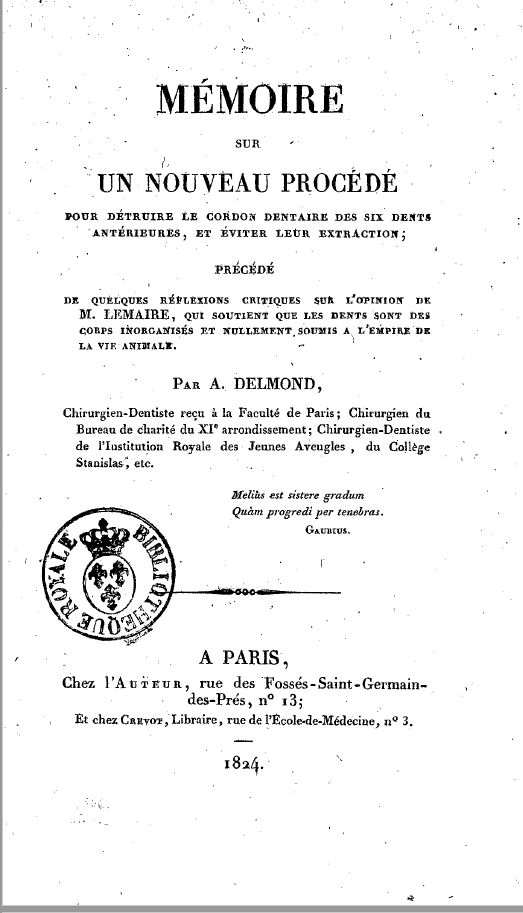
In 1824, Edward Hudson, an Irish dentist who immigrated to America, was the first to perform a treatment that would be vaguely recognizable to modern endodontists when he filled the entire root canal with gold foil. Although he only reported this procedure in 1850, one of his invoices dated 1824 includes the following treatment procedure: “stuffing the cavity of one tooth from the end of its root with gold” (12). An article in 1850 also touted Hudson’s longstanding skill in filling root canals with gold (13). Furthermore, in 1824, when Hudson first filled a root canal with gold, a French dentist named Augustin Delmond (see figure on the right depicting the cover page) described a barbed needle (broach) that could remove the pulp tissue and nerve (14). Here is how M. Desirabode describes Delmond’s method in an English translation of the French work (15): “One of the most specious is the excision and extraction of the dental ganglion; for this operation a small drill is used, which is placed in the cavity of the fang, and, giving the drill a few rotary motions, it is withdrawn with the dental pulp attached.”
In 1836, John Roach Spooner (1794-1838), an American dentist, employed arsenic mixed with sulfate of morphia to devitalize teeth and control the pain associated with pulpitis. It was a procedure published in 1836 by his brother, a dentist, Shearjashub Spooner (1809-1859), under the title Guide to Sound Teeth (16). Even though arsenic became popular over the next century, by 1859, Chapin Harris warned of the side effects (17). Nevertheless, by 1849 the success rate of root canals, at least in experienced hands, was close to 95% (18).
Some of the early methods of using cauterization for devitalization seem barbaric by today’s standards, but clove oil with camphor survived well into the 19th century. It became the basis of the French Bonain’s Solution (equal parts cocaine, camphor, and phenol) (19) and part of the zinc oxide eugenol restoration, which contains either isoeugenol or eugenol, both sedatives.
Eliminating pain and then using the root canal for anchoring may have seemed a logical two-step sequence to the more sophisticated dentist of the 18th century. As experience accumulated in the 19th century about the biology of oral tissues and the importance of innervation, not everyone was as eager to remove the pulp. A small opening at the bottom of a deep carious lesion occasionally exposed bleeding and inflamed but still vital pulp. In such cases, attempts were made to cap and preserve the pulp. The first pulp capping procedure was described in 1756 by a German physician Phillipp Pfaff, who recommended placing a folded concave piece of thin gold, lead, or other metal plates (20), like a sink drain plug, on top of the pulp. Occasionally, gold was replaced by lead or gutta-percha, and in 1873, lactophosphate of lime (21). Pulp capping received a significant boost in 1920 with the introduction of calcium hydroxide (Calxyl) in dentistry (22) and its popularization as a pulpal wound-dressing agent in the 1930s (23). Whenever pulp capping proved unsuccessful, starting in the 1880s, partial pulpectomy became the recommended course of action. Introduced by Allport, it was described by Taft in his then-newly published 1883 edition of Operative Dentistry (24).
But neither pulp capping nor partial pulpectomy was available for those in the middle of the 19th century when root canals were first emerging. By 1859, root canal filling was becoming more common, and it was achieved using successive layers of gold foil, rolled up into slender cones and then condensed into the canal. As a contemporary publication stated, “Roll them on a fine broach in such a manner as to make a cone-shaped block a little longer than the depth of the canal to be filled, and of the same taper” (25). That same year, Jonathan Taft suggested gold foil be replaced with gutta-percha immersed in chloroform (26), and by 1864 the rubber dam was also introduced by a New York dentist, Sanford Christie Barnum (1835-1885).
Improvements in treating an inflamed or infected root canal did not occur in a vacuum. Major advances were achieved in the latter half of the 19th century regarding our understanding of the microbial nature of infection. In fairly quick succession, Ignaz Semmelweis discovered the cause of puerperal fever and how to prevent it (1846), Louis Pasteur demonstrated that diseases are caused by microbes (1864), Joseph Lister introduced antiseptic surgery (1867), Robert Koch formulated his postulates (1888), and Willoughby Miller (1889) demonstrated the microbial nature of dental caries and how infections could spread via the root canal into the periapical area. As a result, Miller suggested that the exposed pulp should be kept as germ-free as possible. This concept was scientifically verified in 1965 (27).
Even before Miller’s 1893 recommendation, there were attempts to remove necrotic tissue and disinfect the root canal. For instance, in 1874, Adolph Witzel (1847-1906), a German dentist, suggested disinfecting the root canal with creosote before filling it (28). Initially, Witzel took the necrotic pulp that was previously exposed to arsenic and morphine and treated it with phenol, followed by removal of any necrotic tissue using barbed files, cleaning, disinfecting, and finally filling the root with iodoform-containing cement. Willoughby Miller, who had just completed his doctoral thesis with Robert Koch in Berlin on the chemo-parasitic theory of caries, suggested thymol cement, zinc oxide with iodoform, and gutta-percha.
Enlargement of root canals was done with hand instruments, just like cavity preparation. They included fine burs that served the same function as rotary instruments do today. They were neither delicate nor flexible enough to do a proper job. In the following decades, root canal treatment improved due to numerous technological and biological discoveries. In addition to the major microbiological advances, the discovery of general anesthesia, better instruments, the foot pedal-operated drill (1871), and the light bulb (1878) all pushed dentistry and root canal therapy toward becoming a more established, albeit not yet fully educated profession. In 1880 in the US, for instance, 3,177 practicing dentists graduated from dental school (29), representing only about a quarter of the 12,314 dentists in the US (30,31).
Two additional developments improved root canal therapy: the accidental discovery of the X-ray by Wilhelm Conrad Roentgen in 1895 and the Gies Report in 1925, which urged all dental educational institutions and disciplines to adopt a scientific basis. By 1896, C. Edmund Kells Jr., a graduate of New York College of Dentistry’s Class of 1878, was the first to use X-rays in dentistry in the US at New Orleans Dental College (32). By today’s standards, it seems rather rudimentary. Films were held by the dentist with bare hands, and radiation exposure, scattered everywhere, lasted 15-20 minutes for each film. There was not even a lead apron for protection. Indeed, Kells developed radiation-induced cancer of his finger, and after many unsuccessful amputations to contain the radiation damage, he committed suicide (33).
During the last forty years, a remarkable surge of technical advances unique to endodontics or of general nature has made endodontic therapy highly predictable with long-term success. First, digital radiography was introduced in 1987. In 1989, rotary endodontics was added, making canal enlargement and debridement far more standardized. (34) Visualizing a tooth and its root canals were made even better with the introduction of Cone Beam Computed Tomography (CBCT) in the late 1990s. CBCT was approved by the FDA in 2001. The most promising and potentially transformative advance in root canal therapy is regenerative endodontics, first proposed in 2004 (35). Several promising research avenues exist for the regenerative potential of pulp-containing stem cells to revascularize teeth and regenerate dentin (36).
The gradual technical difficulties that emerged in treating root canals eventually required a good deal of experience and specialized skills. Finally, in 1943 a group of dentists who primarily focused their practices on root canal therapy formed the American Association of Endodontists, and by the 1950s, they had established advanced education programs. Initially, such programs were only 12-month-long courses. But they gradually grew into two- and three-year programs over the years. In the past 50 years, endodontics has become a specialty recognized around the World.
- 1. Gutmann
- 2. Vigo p.532
- 3. Hoffmann-Axthelm, p. 311.
- 4. Zias
- 5. Hoffmann-Axthelm p.95, p. 120, and 157.
- 6. Vesalius p.45, Fig 1E.
- 7. Fauchard p. 254-255 Fig 33-34.
- 8. Burnet p.274-5
- 9. Hartman p.138
- 10. Faventini, p.46
- 11. Hoffmann Axthelm, p. 51
- 12. ibid p.314, fig 361
- 13. E.T. p.12
- 14. Delmond
- 15. Desirabode, p.177
- 16. Spooner p.116
- 17. Harris p.351
- 18. Ibid p.347
- 19. Bonain
- 20. Pfaff p.124
- 21. Taft (a). p.301
- 22. Hermann (a).
- 23. Ibid (b).
- 24. Taft (a). p. 297.
- 25. Ibid (b). p. 253,
- 26. Ibid (b). p.277
- 27. Kakehashi
- 28. Witzel
- 29. Taft (a). Fig 94, p. 323
- 30. Koch p.69
- 31. US Census Bureau p.760
- 32. Langland p.681
- 33. Raper p.74
- 34. Wildey and Senia
- 35. Banks
- 36. Jung
A brief timeline of endodontics
Ancient Greeks – hot wire cauterization of dental pulp
1st Century – Galen of Pergamon – open (trephane) the pulp and place nitric acid if tooth painful
9-10th c. Rhazes, Albucasis, Avicenna – acid, arsenic, vitriol placed on the pulp to control dental pain
1500 – Martin Ruland the Elder – camphor placed onto the pulp
1543 – Vesalius – dental pulp described
1632 – Johannis Hartmann – camphor oil placed onto the dental pulp
1728 – Fauchard – uses root canal for dowel pin anchoring of carved teeth
1756 – Phillipp Pfaff – first pulp capping with concave shaped gold, lead or other metal
1824 – Edward Hudson – fills the root canal with gold
1824 – Augustin Delmond – barbed needle used to remove nerve
1836 – John Roach Spooner – uses arsenic and sulfate of morphia for inflamed pulp
1859 – Jonathan Taft – root canal enlargement and filling with gutta-percha softened in chloroform
1864 – Sanford C. Barnum – patents rubber dam
1874 – Adolph Witzel – disinfection of root canal – creosote
1880s – Allport suggest partial pulpectomy
1889 – Bonain formulates the “Bonain Solution” (cocaine, phenol, menthol)
1890s – Willoughby Miller – thymol cement, zinc oxide/iodoform gutta-percha root canal filling
1896 – C Edmund Kells – first dental X-ray – Tulane University
1920 – Calcium hydroxide pulp capping
1987 – Digital radiography
1993 – Rotary endodontics*
1990s – Cone Beam Computer Tomography (CBCT) introduced
1995 – Surgical operating microscopy (SOM)
2004 – Regenerative endodontics – proposed
*Rotary endodontics was originally conceived in 1886, but full acceptance of the technique had to wait a century.(Oltramare, 1892).
References and notes on Endodontics
Banks F, Trope M (2004). Revascularization of immature permanent teeth with apical periodontitis new treatment protocol? J Endod. 30:196-200. (First suggestion for pulp regeneration)
Bonain, A. (1899). Sur un nouveau procede d’anesthesie locale pour operer sur la membrane et la caisse du tympan. Bulletin et Mémoires de la Société Française d’otologie, de laryngologie et de Rhinologie, 14:559-562.
Burnet, Thomas (1697). Thesaurus medicinæ practicæ. Ex præstantissimorum medicorum observationibus … summâ diligentiâ collectus … Subsectio tertia. Pro Dentium Dolores, p.274-275. J. Ant. Chouet & Davidis Ritteri, Geneva. [same page, 274 contains multiple recipes for toothache: (a). recipe by Bartholomeo Montagnana, (b). for Peter Foreest for ear infusion with castor oil. Original Latin text is: Auri instillari justi castorei exiguum cum nardo in oleo coctum; (c) for Ambroise Pare – use of egg yolk, turpentine oil and camphor. The original Latin text is: Nil præstantius oleo terebiathinæ chimico cum pulvere Camphoræ; cujus etiam oleum chimicè paratum ett efficacissimum. Item vitellum ovi assi applicatum prodest, ut et theriaca illita. (d). p. 275. Martin Rulandus. The original Latin text is: Guttas aliquot olei Camphoræ gossipio parvo excipio, ac dentis cavernæ applicari jubeo. Martinus Rulandus Curationum empiricarum et historicarum Centuria.], (e). p. 275. https://books.google.com/books?id=mnpmAAAAcAAJ&pg=PA274&lpg=PA274&dq=Bartholomaus+Montagnana&source=bl&ots=BEW0OLr32O&sig=ACfU3U385KuItni9KYdMqF8Ak9QrFUaNsQ&hl=en&sa=X&ved=2ahUKEwiDkc3Ao8XoAhUihHIEHUDQD9AQ6AEwAHoECAgQAQ#v=onepage&q=Bartholomaus%20Montagnana&f=false
Dammaschke T. (2008) The history of direct pulp capping. J Hist Dent. 2008 Spring;56(1):9-23.
Delmond, Augustin (1824). Mémoire sur un nouveau procédé pour détruire le cordon dentaire de six dents antérieures, et éviter leur extraction; précédé de quelques réflexions critiques sur l’opinion de M. Lemaire, qui soutient que les dents sont des corps inorganisés et nullement soumis à l’empire de la vie animale. Paris, 34 p. – A Treatise upon a New Method of destroying the Dental Pulp, Paris.) (https://gallica.bnf.fr/ark:/12148/bpt6k57391598#) (http://www.sudoc.abes.fr/cbs/xslt//DB=2.1/SET=1/TTL=1/SHW?FRST=2). This is a very rare, self-published pamphlet. The method Delmond uses is also described in detail in Antoine Malagou Desirabode (1847). Complete Elements of the Science and Art of the Dentist, 2d. Ed. p. 177. link: https://archive.org/stream/67620160R.nlm.nih.gov/67620160R_djvu.txt Additionally listed in: Illinois State Dental Society (1890). The Transactions of the Illinois State Dental Society at the Twenty sixth Annual Meeting, Volume 26, page 115). One of the most specious is the excision and extraction of the dental ganglion ; for this operation a small drill is used, which is placed in the cavity of the fang, and, giving the drill a few rotary motions, it is withdrawn with the dental pulp attached.” Delmond has described, with care and much detail, this operation. If we have not at hand the proper instrument for this operation, a substitute might be made of iron or platina wire, resembling a single /acies, and which can be readily introduced into the canal of the tooth. By giving the wire a shape resembling a cork-screw, we can with it readily withdraw the dental pulp.“
Desirabode, M. (Antoine-Malagou). (1847). Complete elements of the science and art of the dentist. 2d Ed. p. 177. American Society of Dental Surgeons, Baltimore, MD.
E.T. (1850). Article III. Filling Teeth after the Pulp-Cavity has become Exposed. (Signed E.T.) – , Am J Dental Science, Vol 1, #1, New Series, October., p. 12-16. In 1831, the author (E.T.) witnessed Edward Hudson successfully perform a root canal on a canine.
Fauchard, Pierre (1746). Le Chirurgien Dentiste. 2d Ed. Vol. 2. p.254-258, Fig. 34-35. Chez Pierre-Jean Mariette, Paris. (Dowel pins anchoring prosthesis and carved ivory crowns.)
Faventini, Leonello Victtori Benedicti (1550). Viri in Arte Medica Excellentissimi, Opera, page 46. (for drilling into the tooth and necrotize the pulp). Aliud ad idem, si dens dolens esser perforatus, impleatur foramen cum sequenti mixtura: Rx/ Theriacæ unc.s. pyrethri, nigellae ana drach.s. aquæ vitæ drach.i. misce, et impleatur foramen dentis. “If case of pain perforate the tooth and fill the opening with the following mixture: Theriac, half ounce, chrysanthemum, fennel flower, each, 2 gr., nitric acid ½ a teaspoon, mix and place it into the opening of the tooth.
Another (recipe) for the same: if there is toothache, perforate the tooth and fill the opening with the following mixture: Theriac half ounce, chrysanthemum, fennel flower, each, 2 gr., nitric acid ½ a teaspoon, mix and place it into the opening of the tooth. Based on the composition, the nitric acid would have killed the pulp and remove the pain. This description is the first to indicate a form of root canal treatment.
Glass RI, Zahnder HA (1949) Pulp healing. Journal of Dental Research 28, 97-107.
Grossman LI. (1943). Irrigation of root canals. J Am Dent Assoc, 30: 1915-1917. (Uses solutions of sodium hypochlorite and hydrogen peroxide)
Gutmann, James L. (2008). History of Endodontics In: Ingle’s Endodontics, Chapter 2, p. 36-85.
Harris, Chapin (1849). The Principles and Practice of Dental Surgery, Chapter VI, p.347-348, (Dr. JH Foster has filled 40 teeth with 95% success rate 38 success/40 teeth =95%); p.351 – side effects of arsenic.
Hartmann, Johannis (1632). Praxis Chymiatrica, Odontalgia Cap. LXXIII. Paragraph 4. p. 138. The original Latin text is: Si, qui dolent, dentes cavi sunt, praestans est oleum caryophyllorum, in cujus drachma solutus est camphorae semiscrupulus.( Free translation: For painful cavitated teeth place a mixture of 60 gm of camphor oil and clove oil.) Published in Geneva by Leonardo Chouet.
Hermann BW (1930) (a). Dentinobliteration der Wurzelkanäle nach Behandlung mit Calcium. Zahnärztliche Rundschau 39, 888-9. (Calxyl introduction)
Hermann BW (1936). (b). Biologische Wurzelbehandlung. Frankfurt am Main: Kramer. (Calcium hydroxide as a secondary dentin stimulant).
Hoffmann-Axthelm, Walter, (1981). History of Dentistry. History of Dentistry (Translated from German H.M. Koehler, Chicago:, Quintessence Publishing Co., Inc., p.311 (cauterization of teeth to control dental pain, Hippocrates and Archigenes), p.95, p120, p157 – cauterization and oil for toothache and use of protective tube, Rhazes and Avicenna; p.51 Aztecs used hot resin for dental pulp/pain; p.314, fig 361 – Edward Hudson performed root canal filling with gold in 1824-25; p.311, pulp capping Phillipp Pfaff 1756;
Jung C, Kim S, Sun T et al. (2018). Pulp-dentin regeneration: current approaches and challenges. J. Tissue Regeneration. 10:1-13. (Pulp regeneration).
Kakehashi S., Stanley HR, Fitzgerald RJ. (1965). The effects of surgical exposure of dental pulps in germ-free and conventional laboratory rats. Oral Surg Oral Med Oral Pathol. 1965;20:340-349. doi:10.1016/0030-4220(65)90166-0. (study demonstrating the importance of germ-free environment for a successful endodontic treatment).
Koch, Charles RE. (1910). A profession in which the demand exceeds the supply. Northwestern dental Journal, Volume VIII, Nr. 1. p.69-72. (https://www.google.com/books/edition/Northwestern_Dental_Journal/1mwVAQAAIAAJ?hl=en&gbpv=1&dq=graduates+of+dental+schools+prior+to+1860%3F&pg=PA69&printsec=frontcover) (total number of dental school graduates in 1880 = 3,177, representing 25.8% of the total dentists).
Langland, Olaf E, Fortier, Peter (1972). C Edmund Kells. Oral Surgery, Oral Medicine, Oral Pathology. 34(4):680-689. (C Edmund Kells took the first X ray in 1896).
Miller WD. (1891). The human mouth as a focus of infection. Dent Cosmos, 33: 689-713. (Good review of bacterial nature of many infections in the mouth). link: https://hdl.handle.net/2027/uc1.b3711497?urlappend=%3Bseq=712
Miller WD. (1894). An introduction to the study of the bacteriopathology of the dental pulp. Dent Cosmos, 36:505-528.
Maury, F. (1841). Traité Complet de L’Art du Dentist. 3d Ed. editor Paul Gresset. Paris, Libraire des Science Medicales, De Just Rouvier. (Cauterization needle, Planche XXIV).
Nyborg H (1955) Healing process in the pulp on capping. Acta Odontologica Scandinavica 13,, 1-130. (Pulp capping).
Oltramare, Plotzliche (1892). Exstirpation der Zahnpulpa mittels einer durch die Bohrmaschine in Rotation versetzten Nadel. Dtsch Monatsschr Zahnheilk. 10(9):407–409).
Pfaff, P. (1756). Abhandlung von den Zahnen. Berlin. p.124.
Raper, Howard R (1953). Notes on the early history of radiodontia. Oral Surgery, Oral Medicine, Oral Pathology Volume 6, Issue 1: p 70-81. (C. Edmund Kell’s suicide).
Sedgley, Christine (2004).Root Canal Irrigation Perspective. J Hist. Dent. 52:2:61-64. (review of the history of endodontics).
Spooner S. (1836). Guide to Sound Teeth. New York, Wiley & Long, p. 116 (the use of arsenic and morphium)
Taft, J. (1883) (a).. Practical treatise on operative dentistry. Philadelphia, P. Blakiston Son and Co., 4th Ed.
p. 300-301. (Taft describes the method used by JE Cravens in 1873 of using lacto-phosphate of lime for pulp capping left in place for six weeks.); quoted in Taft, p.297- Allport – partial pulpectomy; p.323, hand instruments devised by Dr. Corydon Palmer for root canal enlarging and filling Fig. 94). p.307 removal of the pulp under local anesthesia using chloroform.
Taft, J (1859) (b).. A practical treatise on operative dentistry. London, Trubner and Co. (p.253 – gold foil root canal filling technique; p.277 use gutta-percha in chloroform instead of gold.
US Census Bureau -1880 – Table XXXIII.-The Number of Persons in the United States Engaged in Each A Total Number of All Persons Occupied. p. 760. (https://www2.census.gov/library/publications/decennial/1880/vol-01-population/1880_v1-20.pdf#) (12,314 dentists). (Number of dentists in 1880).
Vesalius, Andreas (1543). De Humanis Corporis Fabrica Libri Septem. p. 45, Fig 1 E. (Vesalius identified it as “media molaris dentis pars hic delineatur, sinum in dentibus = the middle of the molar delineates a sinus inside the tooth”) Basel. https://babel.hathitrust.org/cgi/pt?id=gri.ark:/13960/t0rr4v32c&view=2up&seq=12&size=125.
Vigo, Giovanni di Rapallo (1560). Practica, De Dolorem Dentium, 1560, Chapter VI, p. 532. on dealing with pain quotes what Galen presumably stated: “Gal. inquit si medicamina dentiu pro eorum doloris sedatione non cotulerint , tunc ad ultimu remediu opus est trafire id est, ad eorum radicitus extirpatione. If everything else fails, open the tooth and remove the nerve”.
Zias, J and Numeroff K (1986). Ancient Dentistry in the Eastern Mediterranean: A Brief Review. Israel Exploration Journal, Vol. 36, No. 1/2, pp. 65-67. (Nabatean skull from 2d c. with incisor having a bronze wire).
Witzel, Adolph (1879). Die antiseptische Behandlung der Pulpakrankheiten des Zahnes :Mit Beiträgen zur Lehre von den Neubildungen in der Pulpa. (disinfecting the root canal with creosote).
Acknowledgments: I am grateful to James Gutmann, Editor of the Journal of History of Dentistry for his helpful discussions and input in this article and to Robert Spielman for editorial help.
Copyright 2023 © HistoryofDentistryandMedicine.com